fbpx

Ultrasound-Guided Biopsies

A breast biopsy is performed to remove tissue from a suspicious area in the breast, which is then examined by a pathologist under a microscope to determine a diagnosis. An ultrasound-guided biopsy is not designed to remove the entire lesion. However, the goal is to remove enough tissue to allow the pathologist to make a diagnosis.

In an ultrasound-guided breast biopsy, the radiologist uses ultrasound imaging to guide the biopsy device to the site of the abnormality.

Ultrasound-Guided Biopsy

When is an Ultrasound-Guided Biopsy needed?

An ultrasound-guided breast biopsy can be performed when a breast ultrasound shows an abnormality such as:

  • A suspicious solid mass
  • Distortion in the structure of the breast tissue

Ultrasound guidance is used to remove tissue samples in these three biopsy procedures:

Core Needle Biopsy (CNB)

Core Needle Biopsy (CNB) uses a large hollow needle to remove one sample of breast tissue per insertion. These biopsies are minimally invasive needle biopsies using ultrasound to show the abnormality to provide guidance for tissue sampling.

During Your Core Needle Biopsy

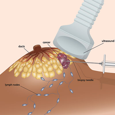
The first part of the procedure will seem much like your original breast ultrasound. You will be positioned lying face-up on the examination table or turned slightly to the side. Prior to your procedure, the skin is cleaned and prepped. Then, numbing medication is injected into the skin and breast tissue. Pressing the transducer to the breast, the radiologist will locate the lesion. A very small incision is made in the skin, just large enough to allow access for the core biopsy needle. The radiologist, constantly monitoring the lesion site with the ultrasound probe, will insert the needle and advance it directly into the mass to obtain the tissue samples. Once the samples are obtained, a clip is placed at the biopsy site to mark where the tissue was sampled. This will be seen on future mammograms. If surgery is needed based on biopsy results, the marker serves as a guide, allowing the radiologist to direct the surgeon to the area that needs to be removed.

Once the tissue samples are obtained, they are submitted to a pathologist for analysis. Results are usually obtained within five working days.

Ultrasound-guided Cyst Aspiration

Ultrasound-guided Cyst Aspiration is a procedure allowing a radiologist to remove fluid from a symptomatic or painful cyst. Using ultrasound imaging, the radiologist can determine the exact location of the cyst. Precisely positioning a needle within the breast, the radiologist then drains the fluid from the cyst. A thinner needle is used for aspiration compared to core biopsy, and clip placement is not needed in most cases.

During Your Ultrasound-Guided Cyst Aspiration

A technologist and radiologist will be working together during your procedure. You will lie on your back or be turned slightly to the side on a padded table. Ultrasound will be used to identify the location of the cyst(s). The radiologist will insert a thin needle into your breast to drain the cyst(s). The fluid from the cyst will drain into a syringe and the cyst will collapse.

Fluid obtained from a cyst aspiration is usually discarded and only in special circumstances sent for laboratory testing. A bandage will be applied over the puncture site.

Cyst Aspirations typically take between 15 to 30 minutes to complete.

Fine Needle Aspiration (FNA)

Fine Needle Aspiration (FNA) is most commonly used to extract sample cells from an enlarged lymph node. The needle used is thinner than a core needle biopsy.

During Your Fine Needle Aspiration

A technologist and radiologist will be working together during your procedure. You will lie on your back or be turned slightly to the side on a padded table. The skin will be cleaned and a small amount of local anesthetic will be applied to numb the area. Ultrasound is used to locate the enlarged lymph node, and cells are obtained through a needle. Samples are then provided to the pathologist for diagnosis.

Fine Needle Aspirations typically take between 15 to 30 minutes to complete.