fbpx

Breast Ultrasound

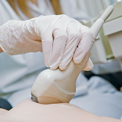
A breast ultrasound uses high-frequency sound waves to make a picture of the tissues inside the breast. The sound waves pass through the breast and bounce back, or echo, from various tissues to form a picture of the internal structures. It is not invasive and involves no radiation or X-rays. The breast ultrasound can show all areas of the breast, including the area closest to the chest wall, which can be hard to study with a mammogram.

At Austin Breast Imaging, we perform both diagnostic and screening breast ultrasounds.

Breast Ultrasound

When is a Breast Ultrasound Needed?

  • To check a lump felt by a doctor during a clinical exam or by a patient on a breast self-exam, after a mammogram is performed.
  • To characterize abnormalities seen on a screening or diagnostic mammogram.
  • To see whether a breast mass is solid or a fluid-filled cyst. Solid masses may require follow-up imaging or biopsy.
  • To look for a cause of spontaneous nipple discharge.
  • To look at the breast tissue in symptomatic women under age 30.
  • To supplement screening mammography in selected patients with dense breast tissue.*
  • In women who are pregnant or should not to be exposed to x-rays.

* Screening breast ultrasounds are performed at Austin Breast Imaging for women with dense breasts. While some centers use a technology called “Automated Whole Breast Ultrasound,” we use the traditional hand-held technique supervised by a radiologist. In our experience, this allows for greater accuracy with fewer false positives.